

Study with the several resources on Docsity

Earn points by helping other students or get them with a premium plan


Prepare for your exams
Study with the several resources on Docsity

Earn points to download
Earn points by helping other students or get them with a premium plan
Community
Ask the community for help and clear up your study doubts
Discover the best universities in your country according to Docsity users
Free resources
Download our free guides on studying techniques, anxiety management strategies, and thesis advice from Docsity tutors
SEUPA THE BIG 4 EXAM QUESTIONS AND CORRECT ANSWERS 100% VERIFIED!!
Typology: Exams
1 / 44

This page cannot be seen from the preview
Don't miss anything!


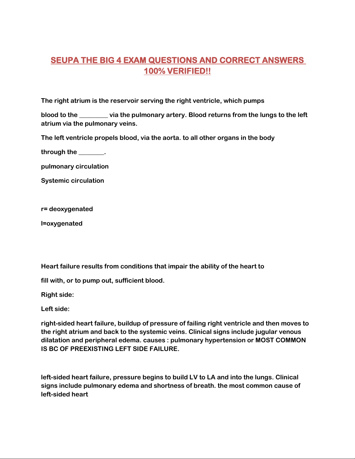
The right atrium is the reservoir serving the right ventricle, which pumps blood to the _________ via the pulmonary artery. Blood returns from the lungs to the left atrium via the pulmonary veins. The left ventricle propels blood, via the aorta. to all other organs in the body through the ________. pulmonary circulation Systemic circulation
r= deoxygenated l=oxygenated
Heart failure results from conditions that impair the ability of the heart to fill with, or to pump out, sufficient blood. Right side: Left side: right-sided heart failure, buildup of pressure of failing right ventricle and then moves to the right atrium and back to the systemic veins. Clinical signs include jugular venous dilatation and peripheral edema. causes : pulmonary hypertension or MOST COMMON IS BC OF PREEXISTING LEFT SIDE FAILURE.
left-sided heart failure, pressure begins to build LV to LA and into the lungs. Clinical signs include pulmonary edema and shortness of breath. the most common cause of left-sided heart
failure is myocardial infarction
ohm's law is pressure and inversely portion to resistance stenosis (severe narrowing) of the aortic give increases resistance to ejection of blood from the left ventricle. According to Ohm's law, left ventricular pressure must increase to maintain sufficient cardiac output. Chronically high left ventricular pressure can result in left ventricular hypertrophy and heart failure.
Poiseuille's law describes the determinants of resistance: R= 8Ln/ pi r^
Radius is the dominant resistance that determines resistance because radius is raised to the fourth power; for example, doubling the vessel radius increases flow by a factor of 16
Rapid fluid infusion ( sized intravenous catheter is applied with Poiseuille law
Compliance describes the distensibility of a structure and is defined as the
heart valve between the left ventricle and the aorta
Coronary ischemia Areas of heart muscle can begin to randomly depolarize. "irritable"· Depolarization of one irritable myocyte rapidly propagates via the all-or- none principle. which can lead to a fatal arrhythmia (ventricular :fibrillation or ventricular tachycardia). Fatal arrhythmias are the most common cause of sudden death during a myocardial infarction
Positive Inotrophy More rapid sequestration of Ca1+ causes faster muscle relaxation
referred to as _____. which occurs with sympathetic nerve
stimulation when the heart rate (HR) is increased and ventricular
ejection time is shorter.
Digitalis (active agent digoxin) is the classic prototype cardiac glycoside
with multiple cardiac effects, one of which is to increase myocardial
contraction via greater loading of intracellular Ca2+ stores.
Although cardiac glycosides can reverse symptoms of heart failure
they art now used with caution due to increased risk of sudden death
Digoxin induces an increase in intracellular sodium that will drive an influx of calcium in the heart and cause an increase in contractility.
atrioventricular (AV) node is the only electrical communication between the atria and the ventricles. (slow for complete contraction) SA node is the normal pacemaker The sinoatrial (SA) node is the normal pacemaker of the heart and the origin of each normal heartbeat
AV node is continuous with the ventricular conducting system, which consists of the atrioventricular bundle (bundle of His), the left and right bundle branches, and the Purkinje fiber system. the first electrical connection with working ventricular muscle occurs through Purkinje fibers.
SA node threshold and pacemaker has no___ The maxim.um membrane potential difference in pacemaker cells is about -60 m V. but there is no stable period of resting membrane potential. ii When the pacemaker potential reaches a threshold voltage of about -40 m V.
four classes of antiatthythmics based on their site of action, class 1 sodium channel blockers; class II, beta blockers; class III, potassium channel blockers; and class IV; calcium channel blockers
SA and AV upstroke depends on _____ where as Ventricles depend on ____. Ca+ NA+
AV node becomes the pacemaker if the SA node fails or transmission to the AV node fails. 1hese patients are in nodal rhythm and typically have resting heart rates of 45-55 beats/ min.
In patients with complete heart block, there is no transmission through the AV node; the His-Purkinje
fibers pace the heart between 20 and 40 beats/min.
i. Slower pacemaker activity in distal parts of the conducting system
allows the heart to continue beating if the SA node fails. However, these
patients have bradycardia (slow HR) and reduced cardiac output
ii For example, sick sinus syndrome occurs when the SA node becomes
fibrotic and loss its ability to spontaneously depolarize. Patients have
bradycardia and symptoms of hypoperfusion such as dizziness, syncope,
weakness, and fatigue
Circular (reentry) conduction occurs when control by the
pacemaker fails and each spread of excitation through the ventricles
triggers the next.
i Reentry is normally prevented because ventricular muscle cells have a
allowing sympathetic tone to continue unchecked
Faster rate is sympathetic stimulation steeper pacemaker potential slope. Effects of autonomic nerve stimulation on nodal action potentials. Parasympathetic (vagaD stimulation reduces the heart rate by hyperpolarizing the nodal cells and reducing the slope of pacemaker potentials. Sympathetic stimulation increases the heart rate by depolarizing the nodal cells and increasing the slope of pacemaker potentials.
P wave represents atrial depolarization
QRS complex represents ventricular depolarization.
T wave ventricular repolarization
Left ventricular hypertrophy is identified if the sum of voltage
deflections for the S wave in lead V1 and the R wave in V5 or 6.
Right ventricular hypertrophy is characterized by an R wave that is larger than the S wave in V
The QT interval correspcmds to the duration of phase 2 of the ventricular action potential. A dangerous form of _______. Class 3 drug prolong phase 2 by blocking ventricular tachycardia called torsade de pointes
frontal lead: transverse plane: 6 limb leads precordial chest leads
bipolar leads = augmented UNIpolar leads = 123 avr avl and avf
negative recording is seen in lead B because the vector is parallel to lead B but directed at Its negative terminal. No net signal Is recorded In lead C because It Is perpendicular to the vector.
Ventricular hypertrophy the thickening (or enlargement) of the myocardium and is a common cause of axis deviation.
i An increased left ventricular mass biases the direction of the net QRS vector toward the left. Left axis deviation can also occur when the abdominal contents physically push the heart up and to the left. such as that which occurs in patients who are obese or pregnant.
right ventricular hypertrophy Usually have a right axis deviation.
When there Is no net recording In a lead (the isoelectric lead), this Indicates that the direction of the QRS Vector Is perpendicular to the lead (aVR In 1hl case).
When the direction of the QRS vector is roughly parallel to a lead, this produces a large net ORS recording (lead Ill in this case). A net negative (downward) recording Indicates that this vector is directed toward the negative terminal of the lead Ill
HR above 100 bpm HR below 60 bpm Tachycardia vs bradycardia
atrial fibrillation no p wave defined , irregular intervals. random depolarization of av node.
In atrial fibrillation, stasis of blood often occurs in the atrial appendages due to the loss of atrial contraction. Anticoagulation therapy may be needed to counter the risk of thromboembolism.
1st degree heart block PR interval >.20 (LONG )
treatment for 3rd degree heart block pacemaker for adequate hr and blood pressure
Pre.mature ventricular contractions (PVC.) occur with no preceding P wave Ventricular fibrillation is fatal within a short period of time due to lack of coordinated ventricular contraction.
The most common cause of death during a myocardial infraction is fatal arrhythmia such as ventricular fibrillation or ventricular tachycardia
Arrhythmias are often caused by electrolyte abnormalities. For example, as plasma K+ concentration increases, hyperkalemia produces the following sequence of cardiotoxic effects: peaked T wave -> prolonged PR interval -> widening of QRS complex -> AV node conduction block -> loss of P wave. merging QRS T wave to make a sine wave pattern degenerate into ventricular fibrillation and asystole.
Blood is pressurized by ventricular contraction and then ejected into the
circulation during systole. Muscle relaxation is followed by ventricular filling during diastole.
Major phases of cardiac cycle a. ventricular diastole (mid- diastole). the atria and ventricles are relaxed. The AV valve are open, and the ventricles passively fill.
b. Atrial systole. a small amount of additional blood is pumped into the ventricles.
c. Isovolumic ventricular contraction. Initial contraction increases
ventricular pressure, closing the AV valves. Blood is pressurized during isovolumic ventricular contraction.
d. Ventricular ejection (systole). semilunar valves open when ventricular
pressures exceed pressures in the aorta and pulmonary artery. Ventricular ejection of blood follows.
e. Isovolumic relaxation. The semilunar valves close when the ventricles relax
and pressure in the ventricles decreases. The AV valves open when pressure in the ventricles decreases below atrial pressure.
f. Atria fill with blood throughout ventricular systole, allowing rapid
ventricular filling at the start of the next diastolic period.
block
A large v wave can be seen in the jugular vein during tricuspid regurgitation RA from RV
Right-sided heart failure results in increased right-sided heart pressures and JVD. In the abdominojugular reflux test the examiner presses on the liver to increase venous return to the right side of the heart JVD is observed if the patient has right ventricular failure.
Left ventricular end-diastolic volume -LVEDV norm: 140mL , Ventricular systole normally ejects a little over one-half of the end-diastolic volume at rest, producing a typical end- systolic volume of about 60- 70 mL.
The stroke volume (SV) the difference between end-diastolic volume and end-systolic volume
Patients with atrial fibrillation lack this atrial kick and may have
inadequate ventricular filling, particularly during physical activity.
- with cardiovascular disease may rely on atrial kick to maintain adequate cardiac output; without it, they may develop heart
failure
Lub is created by the closing of (s1) mitral and tricuspid valves
Dup is created by closing of (s2) semilunar valves high pitch shorter duration
During inspiration A2 and P2 are heard as seperate known as ____ due to deplay of closure of pulmonic valve and earlier closure of the aortic valve Splitting of S.
Paradoxical splitting of S2.. caused by aortic stenosis and left bundle branch block p2 closes first ( pulmonic valve) then aortic